Understanding NF1

Neurofibromatosis type 1 (NF1) is a relatively common genetic condition that is estimated to occur in 1 in 3000 live births worldwide and causes tumours to grow along nerves.1–3

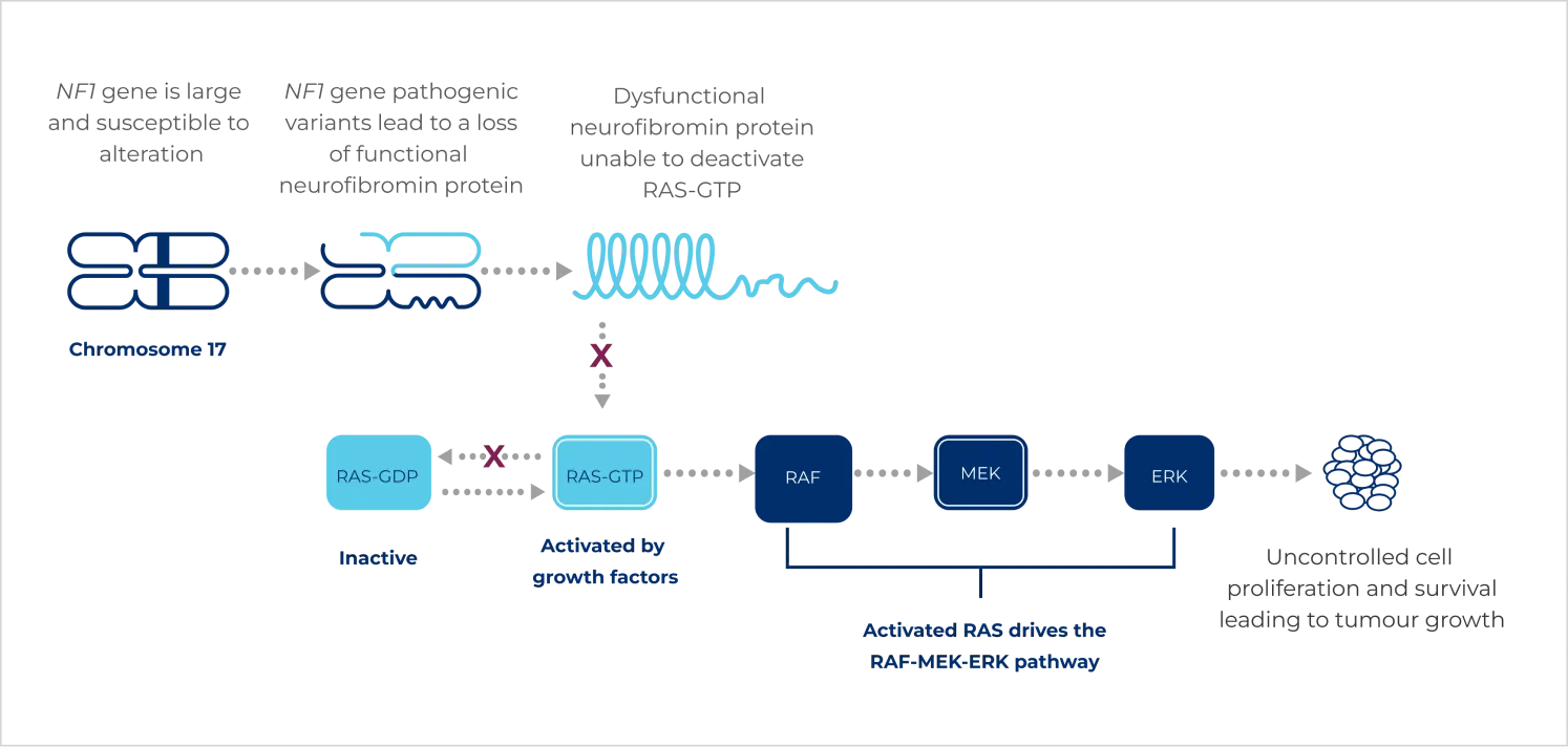
It is an autosomal dominant condition driven by pathogenic variants of the NF1 gene. Pathogenic variants can either be inherited or can occur spontaneously.2 These variants result in the production of dysfunctional or less active neurofibromin, the product of the NF1 gene.4 Neurofibromin is a tumour suppressor protein that regulates the constitutively active rat sarcoma viral oncogene homologue (RAS) protein by keeping it in an inactive form and preventing excessive stimulation of multiple proliferative cellular pathways. Pathogenic variants in the NF1 gene lead to constitutive activation of the RAS- rapidly accelerated fibrosarcoma (RAF)- mitogen-activated protein kinase kinase (MEK)- extracellular signal-regulated kinase (ERK) pathway and uncontrolled growth signalling.4

Adapted from: Yap YS et al. Oncotarget. 2014;5(15):5873–5892.2
Patients with NF1 experience a range of symptoms with varying degrees of severity, which can change over time.2,3 Common symptoms include cutaneous neurofibromas, plexiform neurofibromas (PN), café-au-lait macules, axillary or inguinal freckling, skeletal defects, optic nerve or central nervous system gliomas, as well as neurocognitive dysfunction.1
Common symptoms of NF11,3

Approximately 30–50% patients with NF1 have PN, a tumour of peripheral nerves arising from a bundle of fascicles or a larger nerve plexus.5 PN may remain asymptomatic or can invade surrounding muscle and bone, causing substantial pain and disfigurement.1

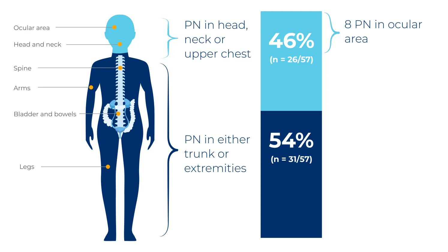
PN are considered congenital and may grow most rapidly in the first decade of life.5 They commonly occur in the head and neck region, trunk or extremities, but can affect nerves between the spinal root and the distal periphery.4,6

PN location from a retrospective review of 41 NF1 patients with 57 distinct PN enrolled in the National Cancer Institute (NCI) natural history study6
Adapted from: Gross AM et al. Neuro Oncol. 2018;20(12):1643–1651.6

The NCI natural history study was a US-based retrospective review of 41 patients with NF1, assessing PN volume and PN-related morbidities in 57 distinct PN over 7 years.6
PN are associated with an increased risk for malignant transformation to malignant peripheral nerve sheath tumours (MPNSTs).1 Patients with NF1 have a cumulative 8–16% lifetime risk of developing MPNST, which usually occurs between the ages of 20 and 35 years.1
Next
ATP, adenosine triphosphate; ERK, extracellular signal-regulated kinase; GDP, guanosine diphosphate; GTP, guanosine triphosphate; MEK, mitogen-activated protein kinase; MPNST, malignant peripheral nerve sheath tumour; NF1, neurofibromatosis type 1; PN, plexiform neurofibroma; RAF, rapidly accelerated fibrosarcoma; RAS, rat sarcoma viral oncogene homologue.
Bergqvist C et al. Neurofibromatosis 1 French national guidelines based on an extensive literature review since 1966. Orphanet J Rare Dis. 2020;15(1):37. Yap YS et al. The NF1 gene revisited – from bench to bedside. Oncotarget. 2014;5(15):5873–5892. NHS. Neurofibromatosis type 1: Symptoms. Available at: https://www.nhs.uk/conditions/neurofibromatosis-type-1/symptoms/ Last accessed: January 2024. Blakeley JO and Plotkin SR. Therapeutic advances for the tumors associated with neurofibromatosis type 1, type 2, and schwannomatosis. Neuro Oncol. 2016;18(5):624–638. Anderson JL et al. Neurofibromatosis type 1. Handb Clin Neurol. 2015;132:75–86. Gross AM et al. Association of plexiform neurofibroma volume changes and development of clinical morbidities in neurofibromatosis 1. Neuro Oncol. 2018;20(12):1643–1651.
advance I am a healthcare professional registered in the EU I am not a
healthcare professional